
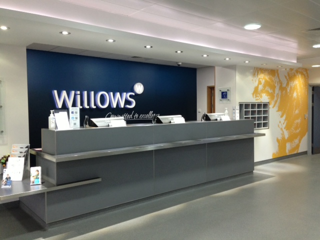
These two weeks I’m placed at Willows referral hospital, which is located in Solihull, West Midlands near Birmingham. Willows offer EMS to final year students, and seeing I passed all my exams my first stop before starting rotations started here.


Willows is a first class hospital for animals that offers: orthopaedics, ophthalmology, anaesthesia & analgesia, internal medicine, soft tissue surgery, neurology and diagnostic imaging as well as having a first opinion small animal clinic. During the 2 week placement I get to follow different cases in all the different disciplines to get a taster of what the specialist field is like. Each patient often receives care from several different teams. I honestly think that these animal hospitals are set up better than many of the human hospitals. I would join the vet of a certain discipline for consults in the morning until around midday. If the patient required surgery, it would be scheduled to undergo diagnostic imaging and then surgery the same day and under the same anaesthetic and either be sent home the same evening or the next day, depending on if the patient had recovered properly.
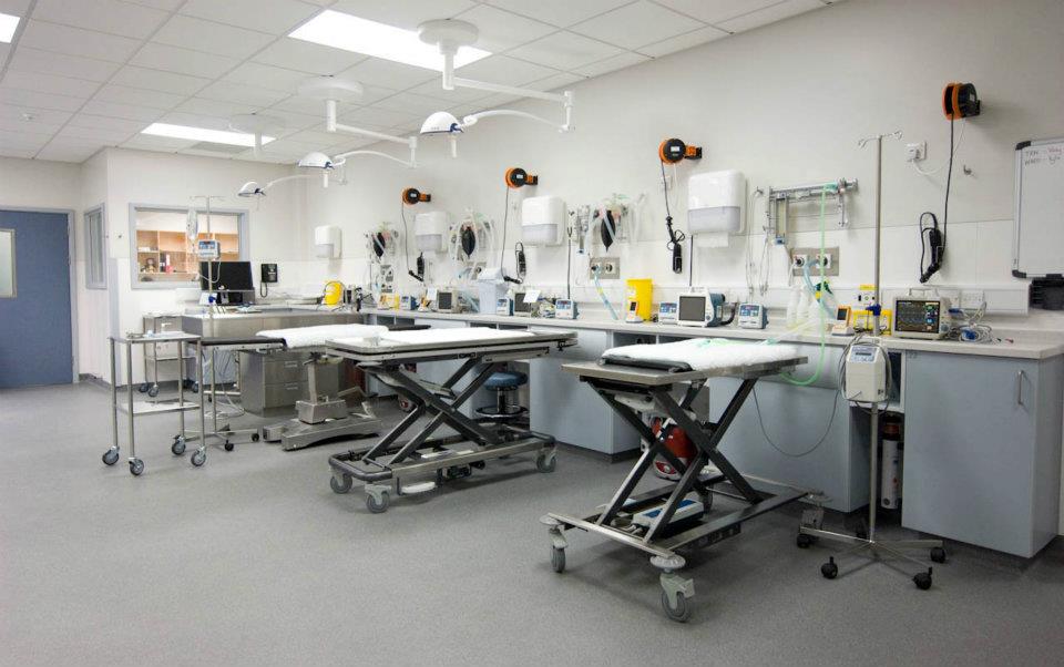
Treatment room
If a patient required special attention the hospital had an ICU ward, where the patients received one to one nursing and checks. The nurses that work at willows knew a lot. There were specific nurses for the different allocated areas, weather it was the dog ward/ cat ward, pharmacy, MRI/ CT nurses, prep room nurses or operating nurses, just like for human hospitals. These nurses had all undergone special training. It seemed to make the hospital run more smoothly.
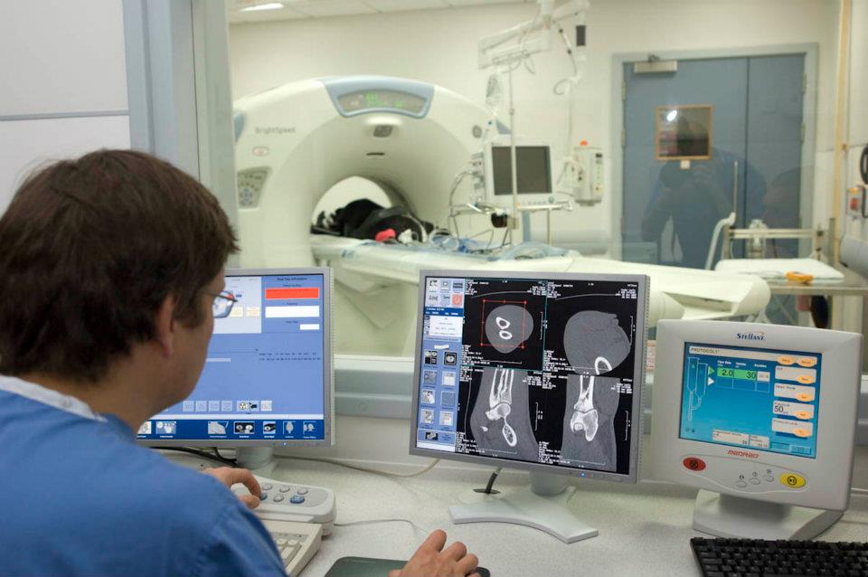
CT
If a patient came from consultations and was now scheduled for a surgery, the patient would get a willows collar with its name written on it (kind of like a hospital wrist band), if the veterinary surgeon was ready to see it in the operating theatre at that point, he would be clipped, the nurse would place an IV Catheter, there would be an anaesthetist vet to write up the drugs it was to get, in the case that it had any special considerations for the anaesthesia e.g: heart murmur, airway problems, geriatric, obese, kidney problems etc. If the animal required x rays for the surgery, the nurses would then take it to the radiography room where they positioned the animals and took the x rays for the vet to have a look at right before surgery. The animal would already be induced by this point, and they had anaesthetic trolleys that they transported the patient to the prep room for the operation.

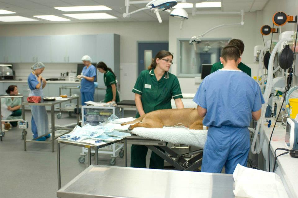
Prep Room
Having these trolleys would prevent the animal being carried, each of the trolleys had isoflurane anaesthesia attached, a breathing system and oxygen so that the patient could be transported sleeping. Once in the prep room all the nursing staff had to wear blue coloured scrubs (vs green for the rest of the hospital) and head caps. The animal was then either induced (if it hadn’t had images taken beforehand) or just prepared, where the surgical site would be cleaned. Once the animal was in the operating theatre the surgical site would be cleaned by a trained nurse again before the site would be draped. All the veterinary surgeons would have to scrub in and put on gowns before operating to ensure that this was as sterile an environment as possible. A nurse would sit and monitor the anaesthesia for the patient throughout the operation and keep records of heart rate, respiratory rate, blood pressure, oxygen etc.
Treatment room
Once the operation was done the animal would be taken out to the treatment room where they would be closely monitored before waking up, given their pain relief and any other medications given before being placed in the kennels. There was a kennel staff who would take patients out for walks as well as making sure the kennels were clean at all times.
My first two days I got to work with Orthopaedics. I was lucky to work with Toby Gemmill and Stephen Kalff consulting on initial referrals or post operative care patients. In the afternoon each day they did TPLO’s which is the abbreviation for tibial plateau levelling osteotomy which is a surgical procedure used to treat cranial crucuate ligament rupture in the knee joints of (often large) dogs. This involved changing the angle of the top of the shin bone (the tibial plateau) by cutting the bone, rotating it, and
  stabilising it in a new position with a plate and screws. Cranial cruciate ligament rupture is one of the most common causes of hind limb lameness in dogs so these surgeons did a lot of this type of surgery.
Wednesday I moved onto Ophthalmology with Mike Rhodes and Carolin Chiwitt. Here I got to see Cataract surgery, keratectomy and cherry eye surgery. Cataracts are an opacity or clouding in the lens in the eye. The lens is normally crystal clear but looks black because the darkness inside the eye. The lens is there to focus light on the sensitive tissue at the back of the eye (retina). Cataracts often form in both eyes and often get worse. It’s more common in older dogs and can be inherited.

phacoemulsification
The only treatment is surgery (but some cataracts are not operable). The surgery is under full general anaesthetic and with a muscle relaxant and then preformed under an operating microscope with tiny instruments. Two small cuts are made in the window of the eye (cornea) and the iris. The eye is filled with a viscoelastic gel to inflate the eye and protect the structures inside. The cataract is then removed though the hole in the capsule using phacoemulsification which is an ultrasound procedure which is also used on human cataracts. After the surgery a special artificial lens is placed where the old lens was, permanent buried deep inside the eye. The surgery wounds are closed with tiny dissolving stitches. Most dogs will actually see on the day after surgery.
Cherry eye is a condition where the third eyelid gland has prolapsed. Dogs have a third eyelid which sweeps back and forth across the surface of the eye protecting and spreading tear film (also called nictitating membrane) which produced 60% of the tear production of the eye. When it prolapses it becomes visible as a pink mass (lump) near the inner corner of the eye. This condition often affects dogs than cats and young animals between 6 to 12mo.

Cherry eye & Pocket technique
Some dogs are predisposed as well: bulldogs, Shih Tzu, Lhasa Apso, Cocker Spaniels, Great Danes and Mastiffs. It can be quite painful and make the eye dry so surgery is recommended, but not to remove it. Pocket technique was carried out at willows where a little pocket is made and the gland is put into it and stitched closed. It sounds like simple thing to do, but this is also done under a microscope with tiny instruments. Ophthalmology is definatly something for the steady handed surgeons.
Friday was Anaesthesia and Analgesia day where I worked with Alessandra Mathis & Anna Bryla. Most of the procedures at Willows will need some kind of sedation, even X rays, Ultrasound scanning or surgery. There are many different types of anaesthetic drugs and many different conditions which require precautions or use of other drugs. Its therefore important to have a good knowledge about patients with often several problems at once, can and cannot get, to prevent drug interactions and other complications.

New week started and I was placed with Soft Tissue surgery with Chris Shales and Stephen Baines. Here I saw surgeries like Tie backs with Laryngeal Paralysis, Porto systemic (liver) shunt surgery and Linear Foreign body removal. Laryngeal paralysis is when there if a functional failure of the larynx opening the vocal cords during inspiration (breathing in). Most cases are older larger breeds like Labrador Retrievers, Golden Retrievers, Weimeraners, Bernese Mountain Dogs, Great Danes etc. Dogs will typically present with a noise when breathing in, coughing, weight loss, reduced exercise tolerance, collapse, reduced tolerance for temperatures, altered phonation (Bark), problems swallowing food/ water, and sudden respiratory distress. A patient doesn’t have to show all of these symptoms. The condition is caused by a dysfunction of one or both the recurrent laryngeal nerves which supply the muscles holding the vocal cords open during breathing. Treatment which is most effective is surgery “Tieback” also called Unilateral Arythenoid Lateralisation (UAL). Which is basically permanently fixing one of the patient’s vocal cords in an open position. 90-95% who have this procedure will improve significantly, but there are some minor complications which can occur as well.

A portal systemic shunt is a blood vessel anomaly that results in the blood from the abdominal organs (small bowel, large bowel, stomach etc) being diverted to the heart or bypassing the liver. This can be a birth defect (congenital port systemic shunt) or acquired if there is a chronic disease. The problem with having a shunt is that nutrients and toxins that should be cleared from the circulation now bypasses the normal portal blood flow to the liver and results in a small liver and build up toxins in the bloodstream which can cause nervous symptoms. The patients often lack necessary substances to give a good supply of energy and therefore have a stunted growth. The surgery I saw was a puppy only 5 mo old. The patients can present with poor muscle development, behavioural abnormalities (walking in circles, disoriented, unresponsive, quiet, staring into space, pressing head into surfaces) and seizures. A portovenogram was done during surgery which is an x ray examination which allows the liver shunt to be accurately visualised during surgery. A vein draining the small bowel is injected with dye which contrasts on the x ray showing the abnormal liver shunt. Surgery is very complex and dangerous for the animal, which involved locating the shunting vessel and closing it to re direct blood through the liver. This might have to be done gradually by placing a cellophane band around the vessel which causes gradual closure over 4 to 6 weeks, giving the liver time to develop without producing an excessively high blood pressure.

Wednesday and Thursday I was on Internal Medicine with Kirsty Roe, Isuru Gajanayake and Amy Lam. Cases with pancreatitis, hypoglycaemic puppies and thrombocytopenia was seen. Pancreatitis occurs when the pancreas becomes inflamed (tender and swollen). It occurs mostly in middle ages to older dogs, breeds like Cocker Spaniels and Terrier breeds. Symptoms ranged from mild signs (reduced appetite) to very severe illness (multiple organ failure). Most commonly it causes lethargy, loss of appetite, vomiting, abdominal pain and diarrhoea. It’s diagnosed with the history along with abdominal pain on examination, but because so much else can cause these symptoms a blood test and ultrasound scan of the abdomen is needed to rule out other conditions.

MRI
Today was unfortunately my last day here at Willows. I was placed with Neurology and Raquel Trevail who had done here residence in Glasgow when I started vet school. We had some follow up cases and also had a patient with Traumatic disc extrusion – slip disc which is when a small fragment of material from the centre of the disc suddenly breaks free and travel through the outer ring of the disc and collides with the delicate spinal cord. Diagnosing this was done with an MRI scan of the animal which I find quite amazing. Willows had a special Diagnostic team who works with Ultrasound, CT and MRI for all the cases that need imaging. Paul Mahoney, Andrew Parry, Lizza Baines and Andrew Tanner are all specialist in Diagnostic imaging to make the best diagnosis and make each sequencing required specially for that animal and that current problem. These two weeks have gone very quickly and I feel very fortunate to have had the opportunity to work with so many great vets & not to forget the nurses. I hope I’ll come back here sometime in the future.
All the best
Annette


Ps: Pictures are from Willows facebook or their website www.willows.uk.net


